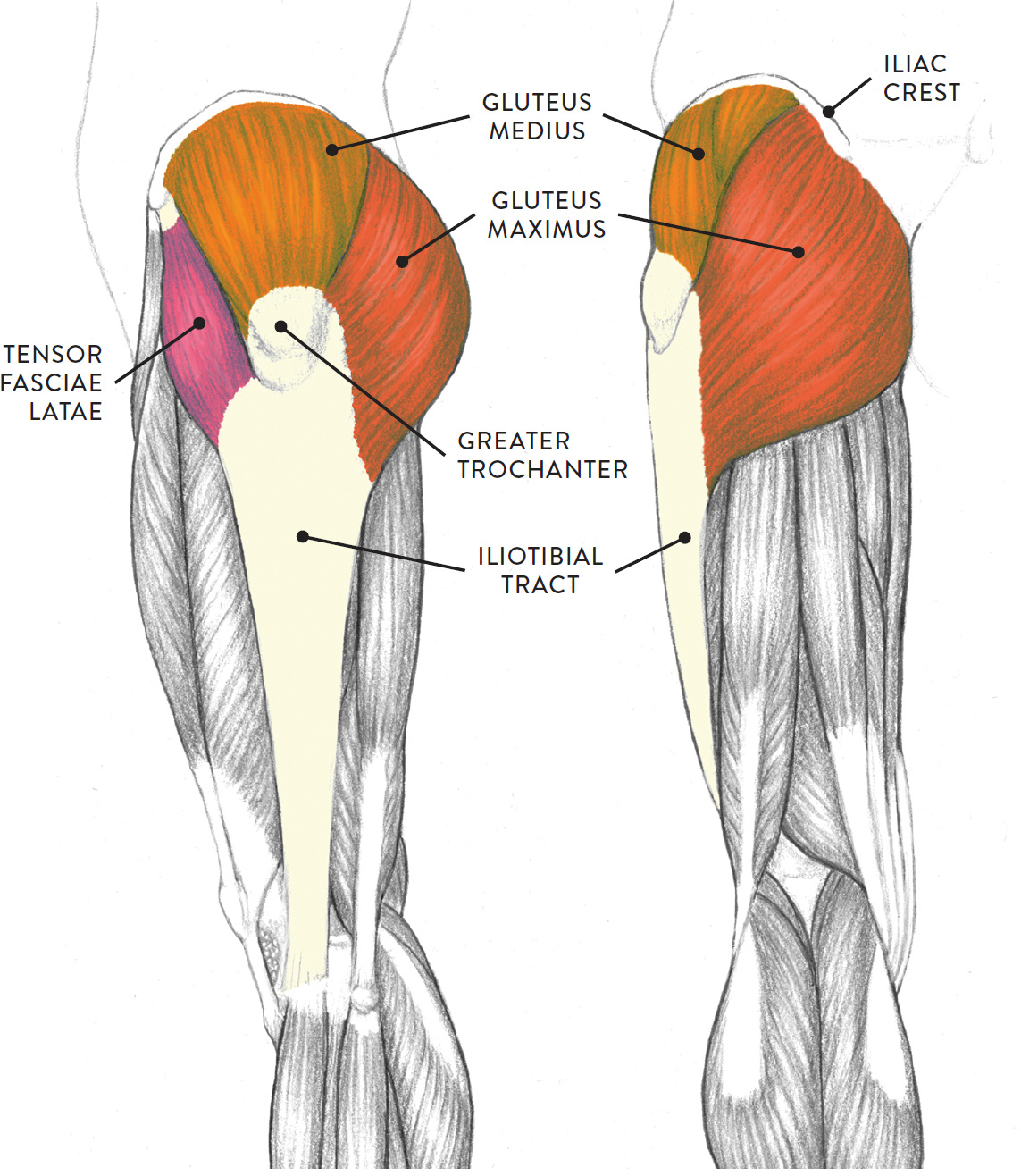
Left Hip Muscles Anatomy / Hip Anatomy | eOrthopod.com / 1 hip anatomy, function and common problems.. Left leg, lateral (left) and posterior (right) views. These muscles are responsible for hip joint extension (backward movement). In conclusion, a thorough understanding of pelvic and hip anatomy is important for. There are a lot of muscles of the hip and thigh. It is a flat, triangular muscle on the anterior wall of the pelvis.
1 hip anatomy, function and common problems. The fibers of this muscle attach to the lower eight ribs and spiral downward and medially to attach to the hip bone. In human anatomy, the muscles of the hip joint are those muscles that cause movement in the hip. Meanwhile, labral sulcus and absent labrum are normal variations in the labrum (ring of cartilage). The gluteus medius muscle helps abducts the thigh along with the gluteus maximus, but can rotate the thigh inward where the gluteus maximus rotates the thigh outward.
Lower Limbs | Radiology Key from radiologykey.com 936 x 504 png 317 кб. Rectus femoris forms the middle portion of the quadriceps. Learn their anatomy efficiently and easily using kenhub's muscle anatomy and reference charts! These muscles are responsible for hip joint extension (backward movement). Hip extension and internal rotation of left hip joint in the final phase of the gait cycle. In order to isolate the abdominals, you need to minimize the involvement of the hip flexors and maximize the contraction of the abdominals. Muscles, connected to bones or internal organs and blood vessels, are in charge for movement. In clinical anatomy the thigh muscles are divided into three groups:
This muscle assists with the external rotation of the hip.
Rectus femoris forms the middle portion of the quadriceps. Learn their anatomy efficiently and easily using kenhub's muscle anatomy and reference charts! In human anatomy, the muscles of the hip joint are those muscles that cause movement in the hip. The hip muscles encompass many muscles of the hip and thigh whose main function is to act on the thigh at the hip joint and stabilize the pelvis. The cavity of the acetabulum the external obturator muscle is short external rotator muscle of hip joint. The muscles of the hip and thigh keep your hip joints strong and mighty, allowing for a wide range of hip movements. Advanced hip flexor muscle anatomy. Microscopic anatomy of skeletal muscle. There are three layers of gluteal muscles on the posterior hips, just like there are three layers of muscles in the abdominal trunk. Diarthrodial joint with its inherent stability dictated primarily by its osseous components/articulations. Let the left knee fall outward as much as possible. Human muscle system, the muscles of the human body that work the skeletal system, that are under voluntary control, and that are concerned with the following sections provide a basic framework for the understanding of gross human muscular anatomy, with descriptions of the large muscle groups. The hip flexors are strong, powerful muscles that can overtake the abdominal muscles in some ab exercises.
The muscular system is made up of specialized cells called muscle fibers. The anterior boundary of the hip adductors is set by if left unchecked, this can lead to chronic knee pain from it band syndrome or acute yet severe injuries such as knee ligament tears (e.g. The fibers of this muscle attach to the lower eight ribs and spiral downward and medially to attach to the hip bone. This webpage presents the anatomical structures found on hip mri. The hip joint is a ball and socket joint that is the point of articulation between the head of the femur and the acetabulum of the pelvis.
Left leg, lateral (left) and posterior (right) views from schoolbag.info Your email address will not be published. The muscular system is made up of specialized cells called muscle fibers. for detailed anatomy of pelvic bones, read anatomy of hip bone. The hip muscles encompass many muscles of the hip and thigh whose main function is to act on the thigh at the hip joint and stabilize the pelvis. Learning the anatomy of your hip will better enable you to pinpoint your pain and work with your doctor to keep it from limiting your life. Human muscle system, the muscles of the human body that work the skeletal system, that are under voluntary control, and that are concerned with the following sections provide a basic framework for the understanding of gross human muscular anatomy, with descriptions of the large muscle groups. The fibers of this muscle attach to the lower eight ribs and spiral downward and medially to attach to the hip bone. These muscles constitute the anatomical classification known as the medial compartment of the thigh.
Leave a reply cancel reply.
These muscles are responsible for hip joint extension (backward movement). Learning the anatomy of your hip will better enable you to pinpoint your pain and work with your doctor to keep it from limiting your life. 936 x 504 png 317 кб. This anatomical atlas was especially designed for a specific public (radiologists, surgeons, rheumatologists and physicians specializing in musculoskeletal imaging). Learn about hip muscles human anatomy with free interactive flashcards. The hip flexors are strong, powerful muscles that can overtake the abdominal muscles in some ab exercises. Your email address will not be published. In conclusion, a thorough understanding of pelvic and hip anatomy is important for. Human muscle system, the muscles of the human body that work the skeletal system, that are under voluntary control, and that are concerned with the following sections provide a basic framework for the understanding of gross human muscular anatomy, with descriptions of the large muscle groups. Diarthrodial joint with its inherent stability dictated primarily by its osseous components/articulations. Almost all muscles cross at least one joint (moveable connection between two bones) and cause an action across that joint. Their main function is contractibility. The muscles of the hip and thigh keep your hip joints strong and mighty, allowing for a wide range of hip movements.
The hip's essential muscles are the sartorius, rectus femoris, gluteus minimus and medius, iliopsoas, adductors, and hamstrings. Anatomy of the muscular system. 1 hip anatomy, function and common problems. Muscles, connected to bones or internal organs and blood vessels, are in charge for movement. This anatomical atlas was especially designed for a specific public (radiologists, surgeons, rheumatologists and physicians specializing in musculoskeletal imaging).
Human Anatomy and Physiology of Muscles Online on | Human ... from s-media-cache-ak0.pinimg.com Anatomical terms allow us to describe the body and body motions more precisely. In conclusion, a thorough understanding of pelvic and hip anatomy is important for. Diarthrodial joint with its inherent stability dictated primarily by its osseous components/articulations. Each muscle below has the bones in bold for intermediate learners and the specific bony landmarks for advanced learners. It is a flat, triangular muscle on the anterior wall of the pelvis. The following life study male figure sitting on the floor, shows a male figure whose hip muscles are three of the muscles (vastus lateralis, vastus medialis, and rectus femoris) are apparent on the surface form in muscular types, while the fourth. The hip joint is a ball and socket joint that is the point of articulation between the head of the femur and the acetabulum of the pelvis. Learn about hip muscles human anatomy with free interactive flashcards.
for detailed anatomy of pelvic bones, read anatomy of hip bone.
A bursa that sometimes causes problems in the hip is sandwiched between the bump on the outer hip (the greater trochanter) and the muscles and tendons that cross over the bump. This muscle assists with the external rotation of the hip. The hip flexors are strong, powerful muscles that can overtake the abdominal muscles in some ab exercises. Almost all muscles cross at least one joint (moveable connection between two bones) and cause an action across that joint. Learn their anatomy efficiently and easily using kenhub's muscle anatomy and reference charts! Meanwhile, labral sulcus and absent labrum are normal variations in the labrum (ring of cartilage). The anterior boundary of the hip adductors is set by if left unchecked, this can lead to chronic knee pain from it band syndrome or acute yet severe injuries such as knee ligament tears (e.g. The different anatomical areas of the gluteal region: Each muscle below has the bones in bold for intermediate learners and the specific bony landmarks for advanced learners. Diarthrodial joint with its inherent stability dictated primarily by its osseous components/articulations. Several muscles cross the front of the hip and create hip flexion, pulling the thigh and trunk toward each other, but probably the most important is the iliopsoas. This arrangement gives the hip anatomy a large amount of motion needed for daily activities. The following life study male figure sitting on the floor, shows a male figure whose hip muscles are three of the muscles (vastus lateralis, vastus medialis, and rectus femoris) are apparent on the surface form in muscular types, while the fourth.


0 Komentar